Maintaining acquisition and analysis data quality
It is important that those running FMRI experiments feed back relevant information to Rhodri Cusack (physics), the radiographers (many issues), the ImagersInterestGroup, the CBU methods group (many analysis and acquisition methods issues) and RhodriCusack (aa analysis in SPM). Anything that will further understanding of the best way to acquire and analyse data is appreciated. If you have a problem, please be constructive, and don't complain when your MR study has failed just because we don't yet fully understand the brain. There will be meetings to discuss data quality every few months open to all using the scanner, as part of the IIG series.If you are designing an experiment, here are some issues you may wish to consider, and a default recommendation for each option.
Contents
Slice order
Brief description
The scanner may acquire slices in an interleaved spatial order (e.g., where 1= bottom slice, 2= next slice up and so on, slices 1, 3, 5... n-1, 2, 4, 6...n) or sequentially, moving always in one direction throughout the acquisition. Both of these may be done in an ascending or descending direction.
Advantages of interleaved over sequential
LESS CROSSTALK: Slices are not perfectly rectangular, and so there is some overlap between spatially adjacent slices. In a sequential acquisition, one slice is acquired just after its neighbour, and the overlapping part is excited twice in quick succession. This may lead to some "saturation" in the overlapping region and resulting loss of signal. In an interleaved acquisition there is a longer interval between the acquisition of spatially adjacent slices.
LESS SLICE TIMING BIAS. See here for a description of the slice timing problem.
Advantages of sequential over interleaved
LESS SPIN HISTORY MOTION ARTEFACT. See here for a description of this problem.
Ascending vs. descending
There is a possibility that if an ascending acquisition is used, the blood flowing up into the brain will be repeatedly excited and saturated. We do not know the degree of this effect.
Recommended default
The default recommendation has changed (3/4/2006): We now suggest that you use a sequential descending sequence (rather than interleaved ascending) unless you plan to reduce the distance factor, ie gap between slices.
Prospective movement correction
Brief description
In contrast to the usual type of FMRI movement correction, which is done offline as part of the preprocessing procedure, the scanner may also perform prospective movement correction (P A C E), in which the scanner measures the position of the head from an EPI acquisition, and then changes the angle and position of the slices to be acquired. The reconstruction (fourier transform from k-space to provide brain images) and movement measurement are done as fast as possible and slice changes are applied to the acquisition that starts 1 TR after the end of the one used to determine the parameters (i.e., lag of 2 TRs).
Possible advantages
- If your subject is going to move gradually throughout the scan, the PACE may keep the slices where you want them on the brain. This might be useful when there is the danger that the brain region of interest will leave the imaging field of view, for example, where only a few slices are being collected, or with a patient that might move substantially.
It might reduce spin history effects. However, as the PACE method involves deriving the motion parameters from the previous EPI, which takes some time, the parameters are adjusted with lag of 1 TR. Any spin history effects will not therefore be corrected in time.
- It is useful for real-time analysis, where you wish to get your results at the same time as acquisition
Possible disadvantages
If there is a large amount of brain activation, it might disrupt the motion estimation procedure, and cause task-related shift in your slice position. This may cause artefactual activation in areas with high distortion (e.g., orbitofrontal), and cause your SPM movement parameters to correlate with your design - see PrinciplesSpatialProcessing.
- If PACE is disabled, we can look for any movement correction problems in the spatial processing phase of the analysis, and try and address it by using different realignment methods, or not doing motion correction. If PACE is enabled, the acquisition itself has changed as a result of the movement parameter estimates, and it is no longer possible to do post-hoc correction.
It may interact with spin history effects
Recommended default
This default has changed (3/4/2006). If you are doing real-time analysis, switch on the prospective correction. If you are not, we recommend that do not use it unless you have a reason to think it is particularly appropriate for your study. Further evaluation is to be performed soon.
Distance factor
Brief description
There is a gap between slices, determined by the "distance factor" which is the size of the gap specified as a proportion of a slice.
Advantages and disadvantages
- A larger gap increases brain coverage and reduces crosstalk (see "Advantages of interleaved over sequential" above for description of crosstalk)
- A very large gap might lead to spin history artefacts with subject movement.
Recommended default
The default distance factor is 25%, with 3 mm slices, giving a gap of 3mmx0.25=0.75mm, and a slice-to-slice distance of 3.75 mm.
Repetition time (TR), field of view & resolution
Brief description
There is a tradeoff between resolution and TR
Advantages and disadvantages
- A faster TR should give higher temporal resolution and possibly greater sensitivity to brief events.
- If you are presenting auditory stimuli, you may wish to introduce a gap between the acqusition of one volume and the start of the next ("sparse imaging") to allow presentation of stimuli in quiet.
- The default recommendation has a short gap (60 ms) at the end, which makes the sound from the sequence slightly discontinuous. If you are interested in time or rhythm, or think this is distracting, you may wish to remove this gap.
- A typical brain is 110 mm in the inferior-superior direction without the cerebellum, and 140 mm wide. A typical sequence 64x64 matrix with 3mm voxels, 32 slices*3.75mm has a field of view of 192 x 120 mm.
- The eyeballs have a high signal variability. There is often a "ghosting" artefact on the EPIs exactly half an image back from the eyeballs, which with flat slices lies somewhere around superior temporal plane or medial temporal regions. It can be helpful to angle your slices upwards at the front by around 30 degrees to avoid these.
How many slices?
The default of 32 slices will cover most of the brain in most subjects. However, if you are also interested in the cerebellum, 36 slices is recommended (this results in a 2.19 second TR). If potentially missing even a few mm of inferior cerebellum (while also acquiring motor cortex) in your biggest-headed subject is a problem, 38 slices may be necessary. Also see note about increasing field of view below.
Recommended default
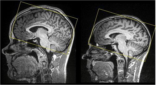
The default CBU sequence has a TR=2s, with 32 slices of 64x64 matrix size, giving a 192x120mm field of view with a resolution of 3x3x3.75 mm. We usually use a slice angle of around 30 degrees to avoid the eyeballs. Typical coverage is shown below on two sample brains.
Note that the person on the left has an unusually large head, and you might want to increase the field of view a little in the ant-post direction , as there will be "wrap around" in your EPIs in this direction, in which the bit of scalp just outside the field of view on the front will appear on the back of your images, and vice-versa.

Slice thickness & angle
Brief description
Because voxels are anisotropic, and different encoding directions are differentially sensitive to field inhomogeneities, the angle of slices can affect the signal loss in regions where there is dropout. The slice thickness also has an effect, as smaller voxels have less of a field change across them for a given field gradient.
Choosing these parameters
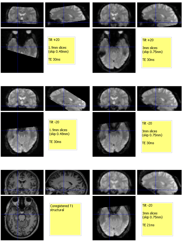
Rik Henson collected the following data to investigate the effect of slice thickness and angle in the frontal lobes. The angles specify the pitch, with positive values corresponding to the slices tipping down towards the front.

A summary of the effects: 1. Less drop-out for thinner slices (compare middle left with middle right panel) - but worse coverage of course
2. Less lateral posterior temporal drop-out when tilting up at front (eg compare top right with middle right panel)
3. ...but more anterior temporal drop-out when tilting up at front (compare where cursor is for top left versus middle left panel)
Note that: i) all volumes contain 32 slices, TR=2.120, each re-shimmed to FoV.
ii) tilts are expressed relative to approximate AC-PC line, which was 12 degrees
Coil
Brief description
Two coils are available - a 12 channel head coil and a single channel transmit/receive coil.
Advantages and disadvantages
- The 12 channel coil has a smaller volume, and so better signal to noise.
- The 12 channel coil gives less homogeneous coverage. This is not usually important for FMRI, but may be important for other applications.
- The single channel coil is used to both transmit and receive RF, whereas the 12 channel coil is used to receive only, with the body coil being used to transmit. If a patient is being scanned or someone else with non-magnetic orthapedic work, it might be preferable to use the single coil as it will excite less widely than the body coil.
Recommended default
The 12 channel coil.
Segment vs. normalise
Brief description
SPM 5 included a new method of performing normalisation, which is simultaneous grey/white segmentation and normalisation.
Advantages and disadvantages
- Segment may perform normalisation more accurately than the standard "normalise option"
However, segment appears to be less robust on our MPRAGE images, either due to the large field of view extending into the neck, or the image inhomogeneity due to the 12 channel coil or parallel imaging. The problem is that the initial affine registration stage mis-converges. One solution is to manually reposition your structural MRI to match the MNI template - as detailed here - http://imaging.mrc-cbu.cam.ac.uk/meg/RepositioningMRIs - though note that if you reposition your structural, you may need to re-coregister your functionals with your new structural position.
Written by RhodriCusack Apr 2006 following meeting on Apr 3. Thanks to all those present for their input.
